A whole-body PSMA PET/CT — the scan that hunts for prostate cancer anywhere in the body. It was ordered because the biopsy showed two more-aggressive features (cribriform and IDC-P), and the team wanted to confirm nothing had spread before choosing treatment. The headline: no evidence of spread.
What lit up
PSMA is a protein on prostate cells; the radioactive tracer sticks to it, so prostate tissue — normal, inflamed, or cancerous — glows on the scan. Two areas inside the prostate took up the tracer, both at low intensity, which is what you'd expect for this grade of cancer.
The known right posterior-lateral tumor lit up, exactly as expected.
A second left-side spot the MRI re-read flagged. Cancer, or just inflammation? Not yet settled — the benign left biopsy favors inflammation.
Where it shows on the scan
These are cropped slices from the PET/CT fusion — the CT gives the grey anatomy, the PET adds the colored tracer signal on top.
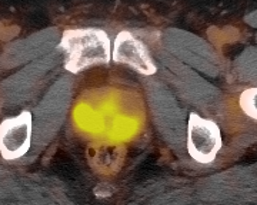
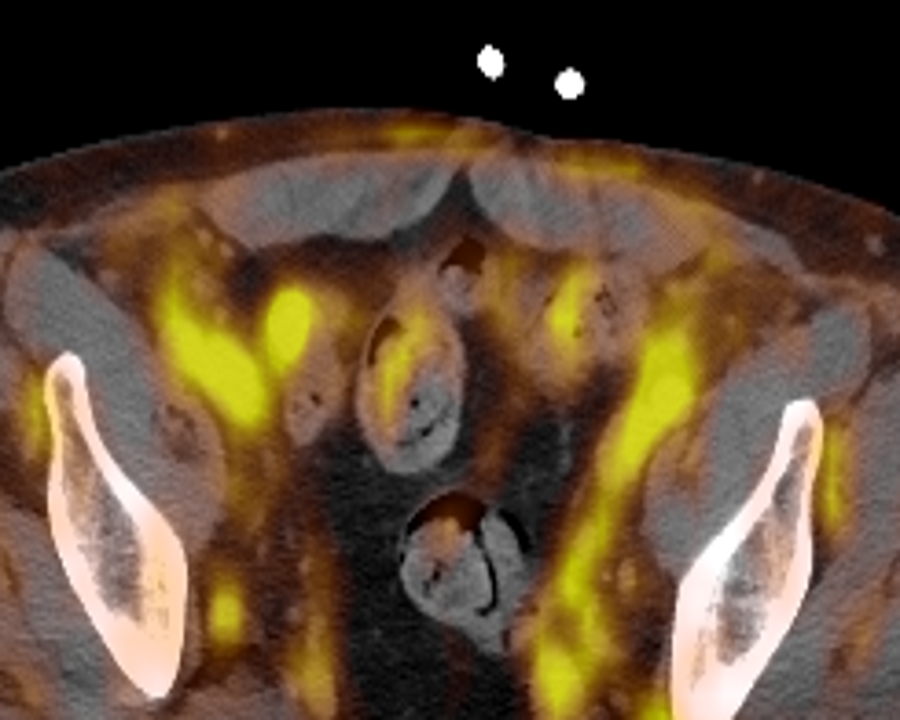
On the first slice, the bright signal low-and-center is the prostate uptake. On the second, look along both pelvic sidewalls: the obturator nodes the Northwell read mentions are small and only mildly avid, so they're subtle — nothing like the bright physiologic activity from bladder and bowel. (MSK's re-read didn't single them out.)
Orientation: axial view — image left = patient right. Tap either image to view it full size.
Signs of spread
The whole point of the scan was to check whether the cancer had traveled. The official impression: no scintigraphic evidence of metastasis.
One nuance, kept honest: the Northwell read notes small, only mildly-avid obturator nodes along the pelvic sidewalls. They don't meet the bar for cancer — and MSK's re-read didn't flag them at all, calling the pelvic nodes clean. Either way the bottom line holds: no evidence of metastasis. And because a PET can't resolve microscopic disease, a faint amount of spread below its detection threshold still can't be fully ruled out — that's a limit of the test, not a finding.
The left-side question
The second left-side focus traces back to MSK's June 10 re-read of the April MRI. Working from the same images, MSK's radiologist flagged a smaller, lower-suspicion spot on the left — PI-RADS 4, about 0.8 cm — the first read to call the left side out as its own target (it's Lesion 2 in the MRI report). The PET then lit that same spot, faintly. The Northwell PET read frames it as inflammation versus a second small cancer, and raised a possible, unconfirmed question of whether it reaches the base of the left seminal vesicle — a question MSK's re-read did not echo, noting no seminal-vesicle involvement at all.
There are real reasons to lean toward inflammation: the left biopsy cores came back benign (a tissue sample is harder to argue with than a scan), the original April read saw nothing on the left, MSK's re-read rated it only lower-suspicion and didn't repeat the seminal-vesicle question, and the PET signal is faint. The scan can't settle it alone — which is exactly why it's first on the care team's list.
On the question it was ordered to answer — has the cancer spread? — the PET came back clean, which keeps both curative paths open: surgery and radiation. The left-side focus and the seminal-vesicle question are what the team settles next. See the treatment options, the MRI report, or the narrative June PET update.
The scan asked one question. The answer was no.