A plain-English read of my prostate MRI — the outside scan from April, re-read by Memorial Sloan Kettering in June. An MRI flags suspicious areas and rates how worrying they look; a biopsy is what actually confirms what's there.
The two areas it flagged
MSK's reading describes two suspicious spots in the prostate's peripheral zone — the outer rim of the gland where most prostate cancers begin. Each gets a PI-RADS score, a 1-to-5 rating of how likely the area is to be clinically significant cancer on imaging.
Right anterior base-to-mid peripheral zone. Mild capsular bulge, no definite extension beyond the gland.
Left posterior base peripheral zone. Mild capsular bulge, no definite extension beyond the gland.
PI-RADS 5 — clinically significant cancer is very likely on imaging. PI-RADS 4 — likely. The score is a probability from the pictures, not a diagnosis; the biopsy and pathology confirm whether cancer is present and its grade.
Where they are on the scan
These are the ADC images from Series 7 — the map where the suspicious tissue shows up darker than the surrounding gland. The arrows are approximate, report-based callouts to orient the eye, not radiologist-drawn contours.
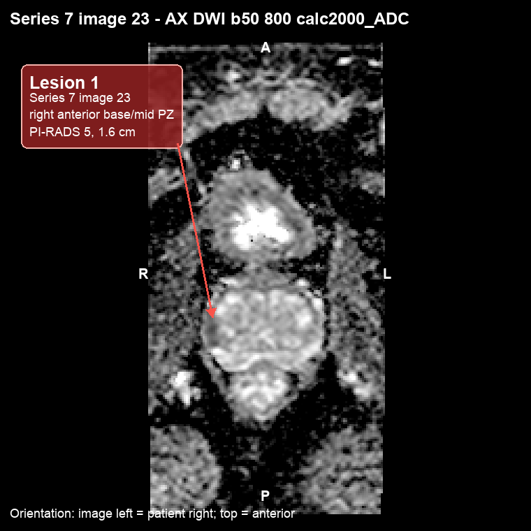
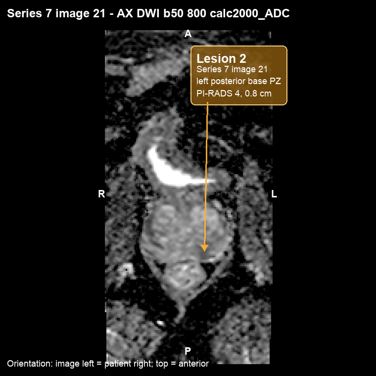
Orientation: image left = patient right; top = anterior. Tap either image to view it full size.
Signs of spread
Beyond the two lesions, the read looks for any sign the disease has moved outside the prostate. On this MRI, each of those checks came back clear:
"No definite extracapsular extension" is the careful phrasing that matters most: both lesions show a mild capsular bulge but nothing the MRI can call as clearly through the wall. Microscopic spread can still sit below what imaging resolves — which is exactly why the surgical pathology read is the tiebreaker.
Two reports, two descriptions
The original Northwell read and the MSK overread use somewhat different anatomic wording. Northwell described a PI-RADS 5 lesion in the right posterior-lateral / anterior horn peripheral zone. MSK described the main lesion as right anterior base-to-mid peripheral zone, and additionally identified the second left-sided lesion.
This page treats MSK as the primary source because it is the later, dedicated overread of the same images.
An MRI strongly suggests, but cannot diagnose. The PI-RADS 5 lesion drove the decision to biopsy, with cores targeted to these exact areas — see the Biopsy & Pathology report for what the tissue actually showed. For the personal narrative around this scan, there's also the original April MRI update.
The picture pointed the way. The biopsy answered it.